Week 7: Neuroscience + Art
This week's topic was a new topic of art that I've never considered before. I never really understood the entire area of what "neuroscience" covers. After doing some more research about what neuroscience is, I found that the topic of neuroscience encompasses all of the scientific disciplines that deal with the nervous system, which is the part of the body that processes the communication between the brain and the rest of the body (“About Neuroscience”).
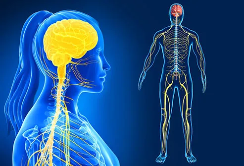 |
| Brain and Nervous System |
The study of the nervous system and brain is a medically applied science, and so it reminded me of the older topic in this class, MedTech + Art. In the video lectures of this week's topic, Franz Joseph Gall was a physician who identified the "grey matter" of the brain as neurons and the "white matter" as ganglia (Vesna, Part 2). It was here where I first noticed that the concepts of "Neuroscience + Art" and "MedTech + Art" were very similar as modern medical technology, such as MRI machines, have given neuroscientists and artists interested with brains a non-surgical technique to access visual representations. Another example of the way the two concepts are associated is through the work of Mark Cohen. In his work, he developed a kind of technology that made the user see the world upside-down. In order to get results from his experiments, he used MRI scans of the users' brains (Cohen).
 |
| Mark Cohen with his vision inverting goggles |
A specific aspect of the connection between neuroscience and art is the project done by Jeff Litchman and Joshua Sanes. They developed a method for identifying specific neurons with the use of fluorescent proteins, which resulted in beautiful images of the brain (Vesna, Part 1). Not only were aesthetic images produced from this project, it also gave a way for scientists to distinguish the neurons from its neighbors. This feature is helpful in tracking the neurons communicating information to other parts of the brain by following the pathways called neural circuits (“Brain Basics: The Life and Death of a Neuron”). From the material provided from this week's topic, I realized that neuroscience and art are dependent on one another.
 |
| Neurons that have fluorescent proteins added to give them colors. (Brainbow) |
Citation:
“About Neuroscience.” Society for Neuroscience, www.sfn.org/about/about-neuroscience.
“Brain Basics: The Life and Death of a Neuron.” National Institute of Neurological Disorders and Stroke, U.S. Department of Health and Human Services, 16 Dec. 2016, www.ninds.nih.gov/disorders/patient-caregiver-education/life-and-death-neuron.
Cohen, Mark. “Neuroscience-Mark Cohen.mov.” YouTube, YouTube, 12 May 2012, www.youtube.com/watch?time_continue=217&v=eDq8uTROeXU.
“Nervous System Pictures: Brain Anatomy and Functions, Nerve Cells.” WebMD, WebMD, www.webmd.com/brain/ss/slideshow-nervous-system-overview.
Vesna, Victoria. “Conscious / Memory (Part 1).” Lecture. 16 Nov 2012. <http://www.youtube.com/watch?feature=player_embedded&v=DLVQIwOn7o8>
Vesna, Victoria. Lecture. “Conscious / Memory (Part 2).” 16 Nov 2012. <http://www.youtube.com/watch?feature=player_embedded&v=Xlg5wXHWZNI>
Weissman, Tamily. “Hippocampus.” Cell, www.cell.com/pictureshow/brainbow?code=cell-site.

I liked how you described the relation between the past material, medical technology and art, and this week's material, neuroscience and art. MRI scans are definitely valuable when used for visually observing the brain without the need for surgical procedures. I also liked your example of using fluorescent proteins to identify specific neurons in the brain. Even though the motive is scientific, the result is a colorful, artistic representation of the brain.
ReplyDelete